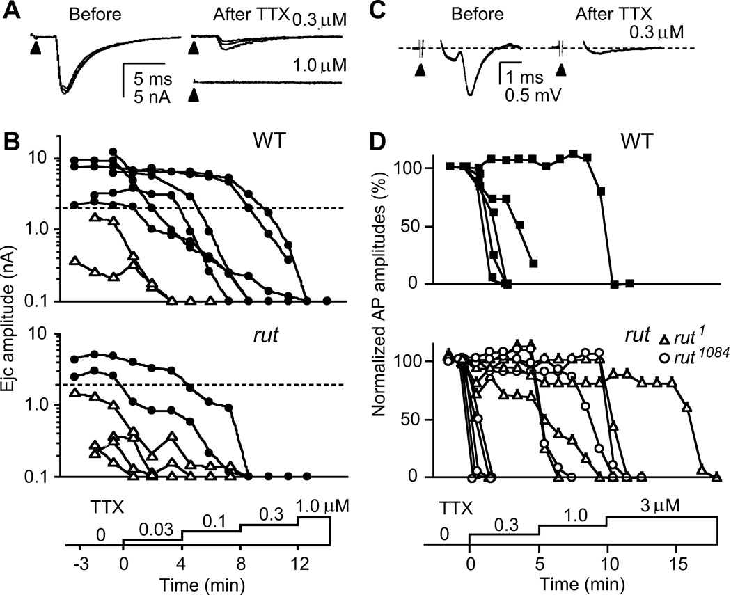
Figure 5.
Sensitivity to TTX paralysis in transmitter release and motor axon action potentials. A. Blockade of nerve-evoked ejcs. Focal ejcs were decreased with a low dose (0.3 µM) and abolished with a high dose (1.0 µM) of TTX (three consecutive traces superimposed). B. Higher sensitivity to TTX paralysis in rut terminals. Ejcs were evoked by 0.1-Hz nerve stimulation while TTX concentrations were increased stepwise (from 0.03 to 1.0 µM). ● and △ indicates ejcs recorded from high- and low-output terminals, respectively. Records from the same bouton were connected with line segments. Note higher TTX sensitivity in low-output terminals. Samples shown reflect the approximate abundance of low-and high-output terminals in rut and WT (Figure 1). C. TTX blockade of axonal compound action potentials. Action potentials were recorded en passant with a suction electrode from the segmental nerve that contains motor axons innervating larval body wall muscles while TTX was applied stepwise at increasing concentrations. Sample traces from WT nerve show compound action potential before and after TTX blockade. D. Overlapping ranges of TTX concentration for action potential blockade of WT and rut motor axons. No significant difference was found between WT and rut alleles in 50 % blockade concentrations (p > 0.05, t-test).

