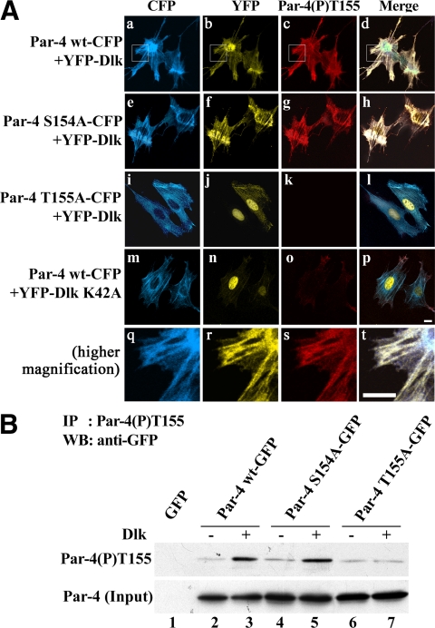
Figure 6.
The Par-4(P)T155 antibody recognizes Par-4 when phosphorylated by Dlk at T155. (A) REF52.2 cells were cotransfected with YFP-Dlk and either Par-4 wt-CFP (a–d), Par-4 S154A-CFP (e–h), or Par-4 T155A-CFP (i–l). As a control, REF52.2 cells were also transfected with Par-4 wt-CFP and the kinase-inactive mutant Dlk K42A (m–p). Twenty-four hours after transfection, the cells were fixed with formaldehyde and stained with the rabbit polyclonal Par-4(P)T155 antibody and anti-rabbit IgG-Cy3 (c, g, k, and o). The phospho-specific Par-4(P)T155 antibody failed to stain cells either coexpressing YFP-Dlk/Par-4 T155A-CFP or Par-4 wt-CFP/YFP-Dlk K42A, suggesting phosphorylation at T155 by Dlk. At higher magnification (q–t) of the insets marked in (a–d) the colocalization of Dlk (r) and phosphorylated Par-4 (s) becomes more evident. Note that both analyses show a slightly punctate staining pattern that matches in the merged image (t). Scale bar, 10 μm. (B) Immunoprecipitation of Par-4 with the phospho-specific antibody Par-4(P)T155. REF52.2 cells were transfected either with GFP-vector, Par-4 wt-GFP, Par-4 S154A-GFP, or Par-4 T155A-GFP alone (lanes 1, 2, 4, and 6, respectively) or cotransfected with the same Par-4 constructs and FLAG-Dlk (lanes 3, 5, and 7). Twenty-four hours after transfection, cell extracts were prepared and subjected to immunoprecipitation with the rabbit polyclonal Par-4(P)T155 antibody. The proteins were separated by SDS-PAGE and analyzed by Western blotting with the monoclonal anti-GFP antibody. Note that in conjunction with Dlk only Par-4 wt-GFP (lane 3) and Par-4 S154A-GFP (lane 5) were precipitated with the anti-Par-4(P)T155 antibody but not Par-4 T155A-GFP (lane 7). The input (bottom) represents 20 μg of whole-cell lysate, which was simultaneously subjected to SDS-PAGE and Western blot analysis with the anti-GFP antibody.

