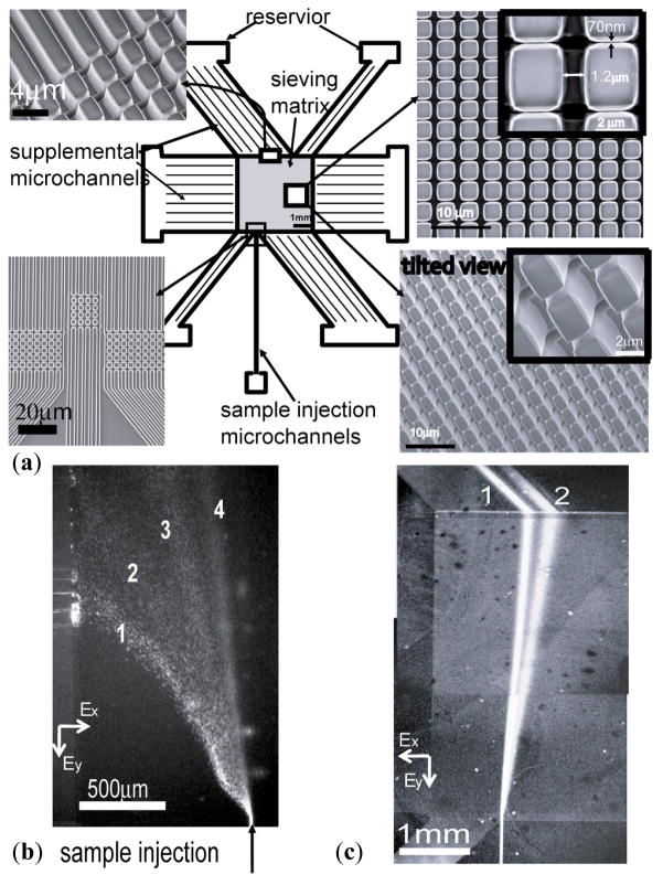
Figure 6.
Continuous fractionation of biomolecules through the two-dimensional anisotropic pillar array device. (a) The device includes a sieving matrix and surrounding microfluidic channels. The pillar array consists of horizontal nanochannels with a width of 70 nm and longitudinal microchannels with a width of 1.2 μm. Supplemental microchannels connecting sieving matrix and reservoirs are 1.5 μm in width. They are all 15 μm deep. (b) Fluorescence micrographs show separation of the mixture of λ-DNA Hind III digest. Electric fields Ex and Ey applied both in horizontal and longitudinal directions in the sieving matrix are 80 V cm−1 and 30 V cm−1, respectively. Band assignment: (1) 23.13 kbp; (2) 9.4 kbp; (3) 6.58 kbp; (4) 4.36 kbp. (c) Fluorescence micrographs showing separation of the mixture of FITC (2) and R-phycoerythrin (1). The nanofilter size is ~40 nm. Ex=250 V cm−1 and Ey=40 V cm−1. It is obtained by combining two fluorescence micrographs taken in the same run but with two different filter sets since they have a different spectrum.

