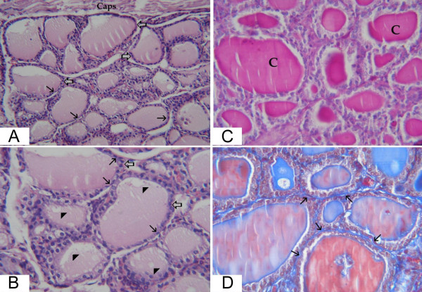
Figure 2.
light microscopic pictures of thyroid gland of group II (ovarictomized rat). (a) large peripheral thyroid follicles (thick arrows) beneath the capsule (caps). The central ones are variable in size with cuboidal and flattened epithelial lining (thin arrows) (Hx&E × 200). (b) higher magnification of the central zone from the previous section showing enlarged follicles (thick arrows) with flat epithelial lining and flattened nuclei of many cells (thin arrows). The colloid demonstrates minimal vacuolization (arrow heads) (Hx&E × 400). (c) follicles distended with large amount of colloid (C) (PAS × 400). (d) increased connective tissue in between the thyroid follicles (thin arrows) (Masson trichrome × 400).

