Fig. 2.
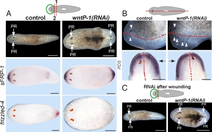
wntP-1 is required for regeneration polarity and anteroposterior patterning. (A) Freshly amputated head fragments (amputation 1) were injected with control or wntP-1 dsRNA for 3 days, amputated again posteriorly on the third day (amputation 2), and allowed to regenerate for 12 days. (Upper) Control head fragments regenerated normally (100%, n = 35), whereas wntP-1(RNAi) animals regenerated a posterior-facing head (26%, n = 77, three experiments). (Lower) In situ hybridizations of control or wntP-1(RNAi) animals at 12 days of regeneration probed with sFRP-1 and frizzled-4 riboprobes. Images are representatives: sFRP-1-stained wntP-1(RNAi), 6 of 24 animals; other panels ≥7 of 7 animals. (B) Intact animals were injected for 2 days and amputated sagittally the following day. (Upper) Uninjected lateral fragments regenerated normally (100%, n = 46), whereas wntP-1(RNAi) fragments regenerated with supernumerary photoreceptors (40%, n = 42) by 10 days. (Lower) in situ hybridizations of laterally regenerating control and wntP-1(RNAi) fragments using a prostaglandin-D synthetase (PDS) riboprobe. Black arrows, newly regenerated side. Extent of PDS in situ hybridization signal was greater on the regenerated side in wntP-1(RNAi) (3 of 5 animals with extra photoreceptors), but not in control animals (7 of 7 animals). Red line, approximate old/new tissue boundary. (C) Day 13 regenerating head fragments that were injected with indicated dsRNAs 1 h after decapitation. Diagrams: RNAi and surgical strategies. PR, photoreceptor. Anterior, left (A, B Upper, C) or top (B Lower). (Scale bars: 200 μm.)

