Fig. 3.
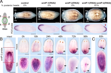
wntP-2 acts with wntP-1 to control regeneration polarity. (A) Diagram depicts RNAi and surgical strategy. Freshly amputated trunk fragments were injected with indicated dsRNA for three consecutive days, amputated near the anterior and posterior wound sites again on the third day, and allowed to regenerate 14 days. wntP-1 and wntP-2 individual injections were mixed with equal amounts of control dsRNA. (Upper) Percentage of animals that regenerated a posterior head. (Lower) In situ hybridizations of regenerating trunk fragments with PDS riboprobe. Red arrow, anterior marker expression in posterior blastema of wntP-1(RNAi); wntP-2(RNAi) double RNAi animals. (B) wntP-2 in situ hybridizations of intact animals and regenerating head and tail fragments at time points (h) after amputation. (B) wntP-2 was detected in an apparent posterior-to-anterior gradient, internally at the anterior pharynx end. New expression of wntP-2 was detected at posterior wounds in regenerating head fragments from 24 h and at progressively anterior locations as regeneration proceeded (p, expression at the new pharynx in a tail fragment). Black arrows, new wntP-2 expression during regeneration (heads) or anterior limit of wntP-2 expression (tails). (C) wntP-2 expression was detected in the posterior of head fragments (solid arrow) but not in the anterior of decapitated fragments at 65 h (empty arrow). PR, photoreceptor. Anterior, left (A) or top (B and C). (Scale bars: 200 μm.)

