
Figure 3. PGC-1α is regulated through post-translational modification.
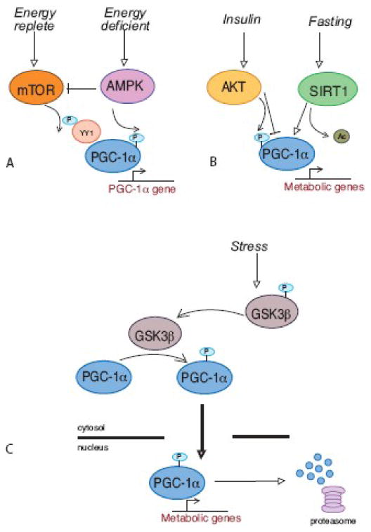
These highly simplified illustrations are intended to convey conceptually how the metabolic impact of any given stimulus can be tailored through alterations in PGC-1α localization, binding partners and gene target specificity. A. Mitochondrial adaptation in response to changes in nutrient availability is achieved through a balance of factors that regulate PGC-1α levels and activity. It is likely that PGC-1α function is not equivalent in each response due to simultaneous regulation of PGC-1α binding partners. B. Tissue specificity plays an important role in the response of PGC-1α to the fed or fasted state. The appropriate response to fasting requires increased glucose output from liver and reduced glucose utilization in skeletal muscle, both of which involve PGC-1α. The use of the same regulator in each tissue ensures a coordinated response. C. PGC-1α localization and stability is regulated in response to stress. Transient activation of mitochondrial metabolism is an early event in the stress response. PGC-1α accumulates in the nucleus and is degraded following transcriptional activation. A strategy of altering PGC-1α protein turnover provides a sensitive and rapid mechanism for regulation of mitochondrial metabolism.