
Figure 5.
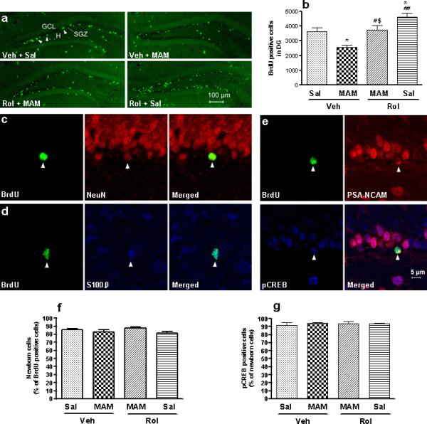
Effects of MAM and/or rolipram (Rol) on BrdU-, PSA-NCAM-, and pCREB-labeled cells in the hippocampal dentate gyrus in mice. (a) Confocal micrographs of BrdU-labeled cells (green) in the dentate gyrus from mice repeatedly treated with vehicle (Veh), MAM, rolipram, or MAM + rolipram. The majority of the BrdU-labeled cells were located in the subgranular zone (SGZ, indicated by arrow). (b) Quantification of BrdU-positive cells following drug treatments. Rolipram increased, while MAM decreased, BrdU-positive cells in the SGZ; the effect of rolipram was reversed by MAM. (c and d) Phenotype of BrdU-positive cells in the dentate gyrus. Confocal micrographs of cells double-labeled for BrdU (green; left panels) and NeuN (red; middle-upper) or S100β (blue; middle-lower). The proportions of neuronal and glial cells (71.4% and 18.5%, respectively) were not altered by any of the treatments. (e) Co-localization of PSA-NCAM and pCREB in developing BrdU-positive cells. Representative confocal micrographs of cells triple-labeled for BrdU, PSA-NCAM (red), and pCREB (blue) in the dentate gyrus. (f) Effects of MAM and/or rolipram on PSA-NCAM-labeled cells, which constituted 84.2% of BrdU-positive cells. (g) Effects of MAM and/or rolipram on pCREB-labeled cells, which constituted 93.4% of PSA-NCAM- and BrdU-positive cells. These percentages were not altered by MAM or rolipram alone or in combination. BrdU (100 mg/kg) was injected (i.p.) once a day on days 10, 12, and 14 of rolipram treatment. Mice were perfused and brain sections were processed 9 d after the last of the three BrdU injections. Values shown are means ± S.E.M of 4−5 mice per group. * p < 0.05 vs Veh; # p < 0.05, ## p < 0.01 vs MAM + Veh; $ p < 0.05 vs Rol + Sal.