
Figure 3. Human dentate gyrus (DG) treated with double-labeling immunocytochemistry for nestin and glial fibrillary acid protein (GFAP) and stained for Nissl substance. The subject was a 57-year-old female control with no medication.
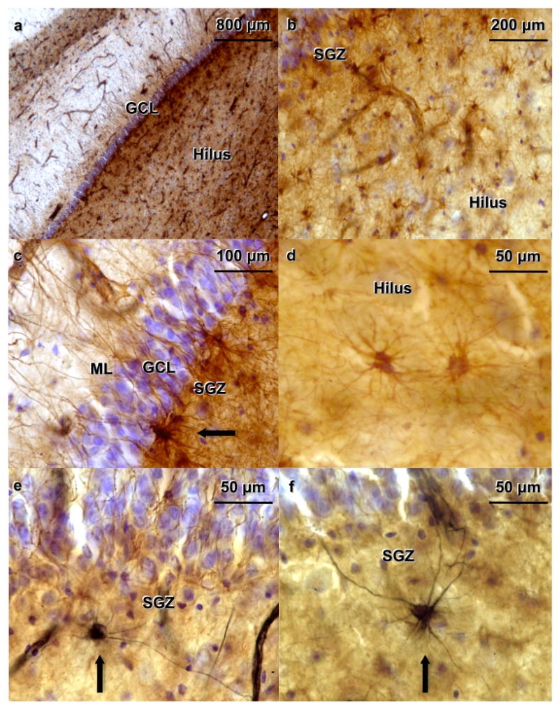
(a) The granular cell layer (GCL) appears in blue, GFAP-immunoreactive (-IR) cells stained with diaminobenzidine (DAB) appear brown. GFAP labels astrocytes in all subregions of the hippocampus (b) GFAP-IR cells are ubiquitously located throughout the hilus. (c) GFAP-IR cells located in the subgranular zone (SGZ) show processes that extend toward the molecular layer (ML), crossing the GCL. A GFAP-IR cell is seen in the SGZ near the lower edge of the picture (arrow). (d) GFAP-IR cells in the hilus at higher magnification. Astrocytes do not stain for nestin, but they do stain for GFAP (e, f) Nestin-IR cells (in black) appear along the SGZ (arrows).