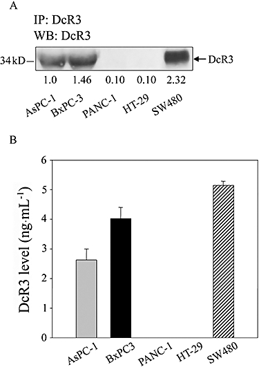
Figure 1.

Immunoprecipitation and ELISA assessment of decoy receptor 3 (DcR3) protein expression in three pancreatic cancer cell lines. (A) Equal amounts of concentrated media from cells were immunoprecipitated (IP) with 1 µg of the DcR3 antibody, followed by immunblot (WB) analysis. (B) Cells (1 × 10⁵) were cultured in 24-well plates. After 24 h, we collected supernatants and measured the DcR3 levels by ELISA. HT-29 or SW480 cells were used as negative and positive controls for DcR3 expression respectively. Data represent the mean ± SEM from five independent experiments.
