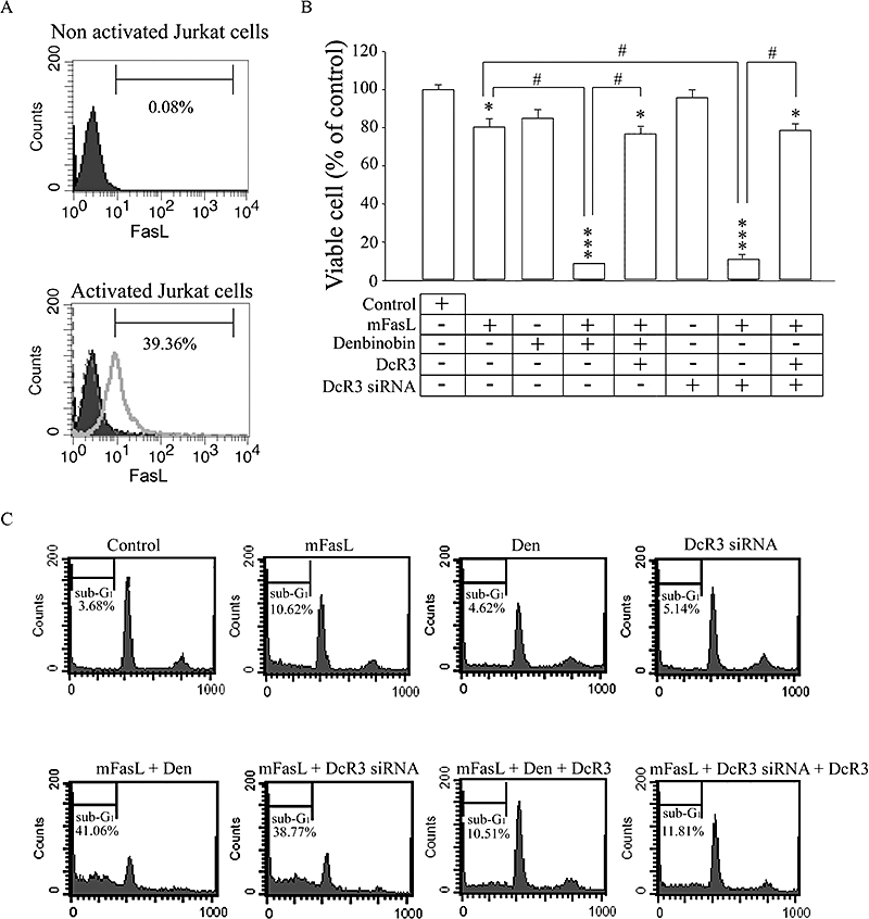
Figure 4.

Denbinobin synergized with membrane-bound Fas ligand (mFasL) on activated Jurkat T cells, in a decoy receptor 3 (DcR3)-dependent manner. (A) Jurkat T cells were cultured in the absence (solid black area) or presence (grey line) of 10 µg·mL−1 of phytohemagglutinin A for 2 h. After incubation, cells were treated with specific FasL antibody to detect the surface expression of FasL by FACScan flow cytometry. Jurkat T cells marked with anti-mouse FITC antibody (dashed grey line) served as the negative control. (B) BxPC-3 cells were seeded onto 24-well plates at a density of 1 × 10⁴ per well. In another group, BxPC-3 cells were transfected with DcR3 siRNA (160 nmol·L−1). After 24 h, medium was replaced with medium containing 3 µmol·L−1 denbinobin and/or 20 ng·mL−1 DcR3/Fc or 5 × 10³ paraformaldehyde-fixed activated Jurkat T cells. MTT assays were performed after 24 h co-culturing. Data represent the mean ± SEM from four experiments. *P < 0.05 and ***P < 0.001 as compared with the control group respectively. #P < 0.01 as compared with different groups as indicated. (C) BxPC-3 cells were treated with vehicle (control), 3 µmol·L−1 denbinobin (Den) or transfected with 160 nmol·L−1 DcR3 siRNA as indicated; these cells were then co-cultured with activated Jurkat cells (as a source of mFasL) for 24 h. Analysis of DNA content was performed by a FACScan flow cytometer. Sub-G1 phase was indicative of apoptosis. Three independent experiments were performed.
