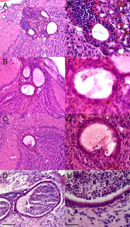
Figure 3.
H&E stained sections showing the morphology characteristics of OB grafts at 30 days. At low magnification (A–D) the formation of multiple vesicles can be observed. At higher magnification (E–H) a multilayered epithelium surrounds the vesicles and ciliated cells can be seen at the surface. Sections shown in A&E are from M1; B&F, M2; C&G, M3; D&H, M6. The OB graft for M5 is shown in Figures 2&4. Scale bar =100um (A–D), 30um (E–H).

