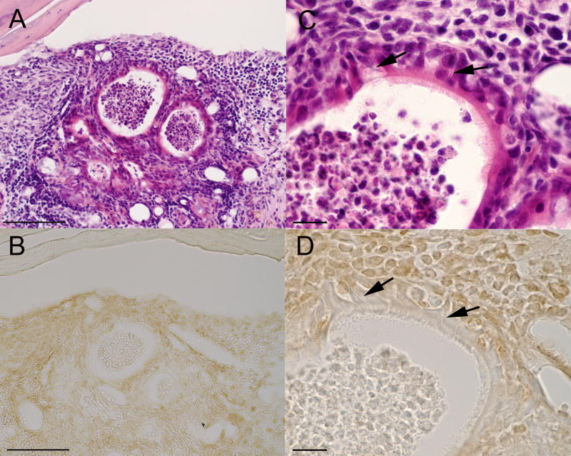
Figure 4.
Histological sections from OB graft site showing the location and morphology of ciliated and neuronal cell types similar to that found in the normal olfactory epithelium. All sections are from the same OB graft site for M5. (A&C) low and high magnification of the same H&E section (also shown in Figures 1B and 2A&B). (B&D) Section cut from same tissue block and immunostained for the Olfactory Marker Protein (OMP). At high magnification (C&D) ciliated epithelial cells can been seen along the surface of the epithelium. Cells with elongated processes (arrows) are similar in morphology to the bipolar olfactory neurons with dendrites extending to the epithelial surface. Scale bar = 100um (A&B), 15um (C&D).

