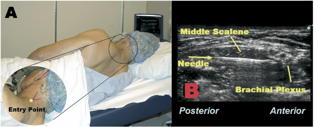
FIGURE 1.
A, The patient is placed in the right lateral decubitus position. The junction of the levator scapulae and trapezius muscles is identified by the “V.” B, A 1 7-gauge Tuohy-tip needle is directed under in-plane ultrasound guidance through the middle scalene muscle toward the brachial plexus.

