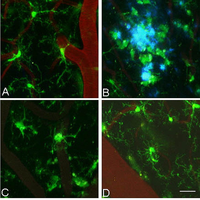
Figure 1.
Microglia have altered morphology and cluster around amyloid plaques in PDAPP+/−;CX3CR1/GFP+/− mice. Three-dimensional reconstructed z-series stack images taken of cortical microglia in (A) PDAPP+/−;CX3CR1/GFP+/− mice at 3.5 months of age (in the absence of plaques), in (B) 14-month-old PDAPP+/−;CX3CR1/GFP+/− mice around amyloid plaques (blue), in (C) 14-month-old PDAPP+/−;CX3CR1/GFP+/− mice in areas lacking plaques, and in (D) 5-month-old PDAPP−/−;CX3CR1/GFP+/− mice. GFP-labeled microglia are green. Vessels are labeled with Texas Red dextran. Amyloid fluoresces blue after injection of methoxy-XO4. Scale bar, 20 μm.

