Figure 7.
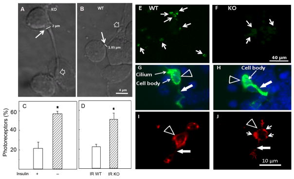
Abnormal development and differentiation of retina photoreceptors from IR knockout mice. Confocal Nomarsky micrographs of retina photoreceptors from IR deleted (A) and wild type (B) mouse retinas grown in serum-free-chemically defined media for 5 days, showing the increase in width of axonal cones (thin arrow in A, B) from 1.05 μm in WT to 2 μm in KO. Percentage of photoreceptors having wider axonal cones in cultures with (+) or without (−) insulin (C) and in cultures obtained from wild-type (IR WT) or knockout (IR KO) mice retinas (D). Confocal fluorescence micrographs of cultures from wild type (E, G, I) and knockout retinas (F,H,J) were labeled with an antibody against IRβ (green staining in E, F), and fluorescence micrographs of cultures double-labeled with DAPI (blue staining) and an anti-opsin antibody (G, H) or with rhodamine phalloidin (red staining in I, J) to examine the organization of F-actin. Photoreceptors of wild type and IR deleted mice showed intense opsin labeling (G, H), even in their neurites (white arrows). Note that in the wild type cultures, IRβ occurrence was conspicuous (E), while in IR-deleted cultures it was barely visible (F). Also note the abnormal accumulation of phalloidin in the cell body (short arrows in J) and neurite (white arrow in J) of IR-deleted photoreceptors. Statistical significance was determined by Student’s two-tailed t-test.*Statistical significant differences compared to controls (p<0.05).

