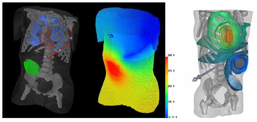
Figure 1.
Left: Hexahedral mesh finite element model of a 2-year old torso, which has been segmented from a CT scan. The heart, lungs, skeleton and body surface are visible, as are an abdominally placed ICD generator and a long subcutaneous ICD lead tracking in the left infra-axillary area. Center: Body surface voltage potentials predicted by FEM for this configuration. Right: A computation of defibrillation fields in an improved torso model consisting of 2 million tetrahedral elements. The ICD can is located in the abdomen and the electrode over the sternum.

