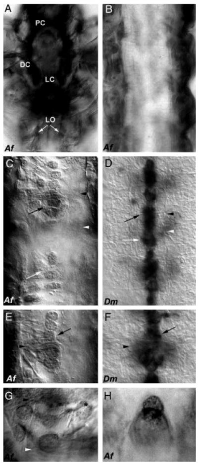
Fig. 2.

Comparison of Netrin central nervous systems (CNS) expression in Artemia and Drosophila. Comparable with the fly, expression of afrNet is found in the Artemia brain during early development (L3 shown in (A); PC, protocerebrum; DC, deutocerebrum; LC, labral commissure). afrNet can be detected on longitudinal (LO marked by white arrows in (A)) axons before commissural axon formation. By stage L6, the longitudinal axon tracts have thickened, and afrNet expression can be detected on many axons (B). Once all of the segments have been generated, afrNet can still be detected on longitudinal axons (dark staining along both sides of trunk in C and E), but can also be detected in midline cells in thoracic segments (C, E, G, H) and on commissural axons (C, E). This midline and commissural axon staining is much less intense than staining on the longitudinal axons, which is also observed in these preparations. Midline expression is comparable with that of stage 13 Drosophila (D, F), though it occurs at a relatively later time point in Artemia. Note that axons are viewed by Nomarski optics only in Drosophila, which are stained through in situ hybridization with a netA RNA probe that labels only the cell bodies (D, F). By contrast, the afrNet antibody marks both cell bodies and axons. Note that one Artemia segment is shown in (C), but three segments are shown in the more compact Drosophila CNS in (D). Black arrowheads mark the anterior commissures, and white arrowheads mark the posterior commissures in (C–G). Black arrows mark the midline glia in (C–F). White arrows mark clusters of cells posterior to the commissures (C, D). Higher magnification views of midline glia encircling the anterior commissure are shown (Artemia in (E), Drosophila in (F)). Net-positive axons in Artemia (G) may correspond to the VUM (white asterisk) and MP (black asterisk) axons. A higher magnification view of posterior afrNet-expressing cells, which may correspond to the MNB cluster in Artemia, is shown in (H). Anterior is oriented up in all figures. Af, Artemia franciscana; Dm, Drosophila melanogaster.
