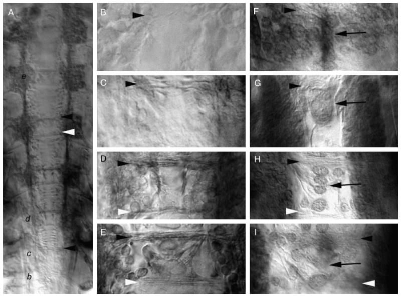
Fig. 3.

Midline expression of Artemia franciscana netrin (afrNet) coincides with formation of the commissural axon tracts. Axonogenesis in Artemia was followed with anti-acetylated tubulin staining (A–E). The temporal gradient of central nervous systems (CNS) development is evident in the L8 animal shown in (A), where commissure formation is initiating in the most posterior segment (marked with black letter b, which is magnified in panel (B). Commissure formation has progressed further in more anterior segments which are marked in panel (A) by the black italicized letters c, d, and e and magnified in the corresponding panels (C, D, and E). Accumulation of afrNet is shown at corresponding stages of development in (F–I). Segments (B) and (F), (C) and (G, D and H, and E and I) are at comparable stages of development. Midline afrNet-positive cells are detected before and during formation of the anterior and posterior commissures (F–I). At later stages of CNS development, afrNet is detected on commissural axons (H and I). These data support a role for afrNet in commissure formation. In this figure, anterior commissures are marked with black arrowheads, and posterior commissures are marked with white arrowheads in various panels. Midline Net-positive cells are marked by black arrows. Anterior is oriented up in all figures.
