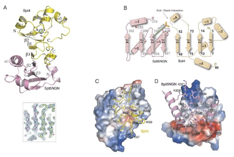
Figure 2. Architecture of Spt4-Spt5NGN complex.

(A) Ribbon diagram of the structure of Spt4-Spt5NGN complex. The Spt4 and Spt5 NGN domains are shown in yellow and pink, respectively; the Zn atom is shown as a blue sphere. The central β-sheet is shown by the 2mfo-Dfc map calculated with refmac coefficients contoured at 2σ level. (B) Topology of the secondary structure elements. Residue numbers and the N and C termini of Spt4 and Spt5NGN are indicated. (C, D) Views of the Spt4-Spt5NGN interface. C, Surface of Spt5NGN is shown and colored according to electrostatic, interacting residues in Spt4 are labeled and shown as yellow sticks; D, Surface of Spt4 and interacting residues in Spt5NGN are shown, interacting residues in Spt5NGN are labeled and shown as pink sticks.
