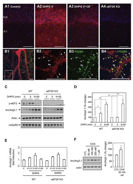
Figure 5. Rapid Induction of Arc/Arg3.1 by Group I mGluRs Is Dependent on eEF2K.
(A) Hippocampal slices were prepared from WT and eEF2K KO mice and were stimulated with DHPG for 5 min. phospho-eEF2 (p-eEF2, red) in area CA1 was increased by DHPG within 5 min and declined by 30 min following washout. Specificity of phospho-eEF2 was confirmed by staining of eEF2K KO slices. s.p., stratum pyramidal; s.r., stratum radiatum.
(B) Cultured hippocampal neurons were treated with DHPG for 5 min and stained with phospho-eEF2 (red) and PSD95 (green) antibodies on DIV14. phospho-eEF2 showed punctal distribution in dendritic spines and dendritic shafts. phospho-eEF2 in spines colocalized with PSD95 (arrows). (B2), (B3), and (B4) are enlarged images of the rectangular region of (B1).
(C and D) mGluR-dependent rapid synthesis of Arc/Arg3.1 is absent in eEF2K KO neurons. Neurons from the forebrains of WT or eEF2K KO mice were cultured for DIV14 and treated with DHPG (50 μM, 5 min). Phosphorylation of eEF2 was undetectable in eEF2K KO neurons. No difference in the level of mGluR5 was observed between WT and eEF2K KO neurons. An arrowhead indicates a non-specific band. p values were obtained by paired t test comparing basal and drug-treated levels. p values for comparison of WT and eEF2K KO mice were obtained by Student’s t test. *p < 0.05, **p < 0.01, n = 8. Error bars are SEM.
(E) Arc/Arg3.1 mRNA expression is not altered in eEF2K KO neurons. The level of Arc/Arg3.1 mRNA was measured in WT and eEF2K KO neurons following the stimulation with DHPG.
(F) Low-dose cycloheximide (CHX) increases Arc/Arg3.1 protein expression. Cultured eEF2K KO neurons were treated with indicated doses of CHX for 10 min. *p < 0.05, n = 8.

