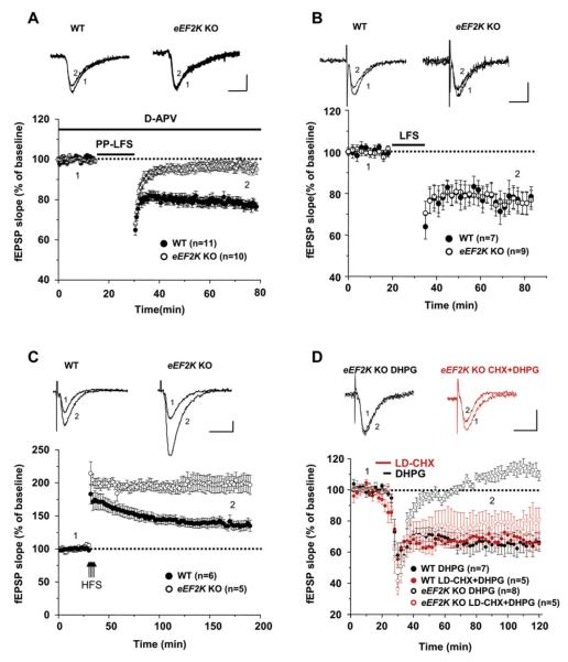
Figure 6. mGluR-LTD Is Impaired in Hippocampal Slices Derived from eEF2K KO Mice.
fEPSPs were recorded in the hippocampal CA1 region of slices derived from eEF2K KO mice and compared to WT littermate controls.
(A) Time course of the change in fEPSP slope produced by paired-pulse low-frequency stimulation (PP-LFS: at 1 Hz, 50 ms interstimulus interval, for 15 min) in the presence of D-APV (50 μM). LTD of WT mice was 77.0% ± 2.1% of baseline at t = 75 min (n = 13). In eEF2K KO mice, fEPSPs were 97.5% ± 2.4% of baseline t = 75 min (n = 15) (p < 0.0001).
(B) Time course of the change in fEPSP slope by low-frequency stimulation (LFS: 1 Hz for 15 min). This form of NMDAR-dependent LTD was not altered in eEF2K KO hippocampal slices (72.7% ± 2.2% of baseline at t = 75 min, n = 9) compared to WT (73.1% ± 3.4% of baseline at t = 75 min, n = 7) (p > 0.5).
(C) Late-phase of LTP was induced by four stimulus trains (100 Hz each) with an intertrain interval of 3 s. In WT, fEPSPs were increased to 171.5% ± 13.4% of baseline immediately after stimulation (t = 30 min) and were sustained at the level of 138.4% ± 7.7% of baseline at t = 175 min (n = 6). However, in eEF2K KO, the initial LTP (204.6% ± 8.9% of baseline at t = 30 min) was maintained for 3 hr after stimulation (200.1% ± 11.9% of baseline at t = 175 min, n = 5). LTP was significantly greater in slices derived from eEF2K KO mice compared to those from WT mice at this time point (p < 0.005).
(D) Average time course of the change in fEPSP slope induced by DHPG (50 μM, for 5 min). LTD of WT mice was 64.7% ± 5.2% of baseline at t = 90 min (n = 7). In eEF2K KO mice, LTD was significantly impaired (108.7% ± 3.6% of baseline at t = 90 min, n = 8). Treatment with low-dose cycloheximide (LD-CHX, 50–75 nM) for 10 min starting from 5 min prior to DHPG restored DHPG-LTD in eEF2K KO (75.7% ± 7.4%, n = 5). In WT mice, treatment with LD-CHX did not alter the expression of LTD (69.0% ± 2.6%, n = 5). p < 0.001 when eEF2K KO DHPG only was compared to eEF2K KO LD-CHX + DHPG, WT DHPG only, or WT LD-CHX + DHPG. Scale bars, 0.5 mV/10 ms.

