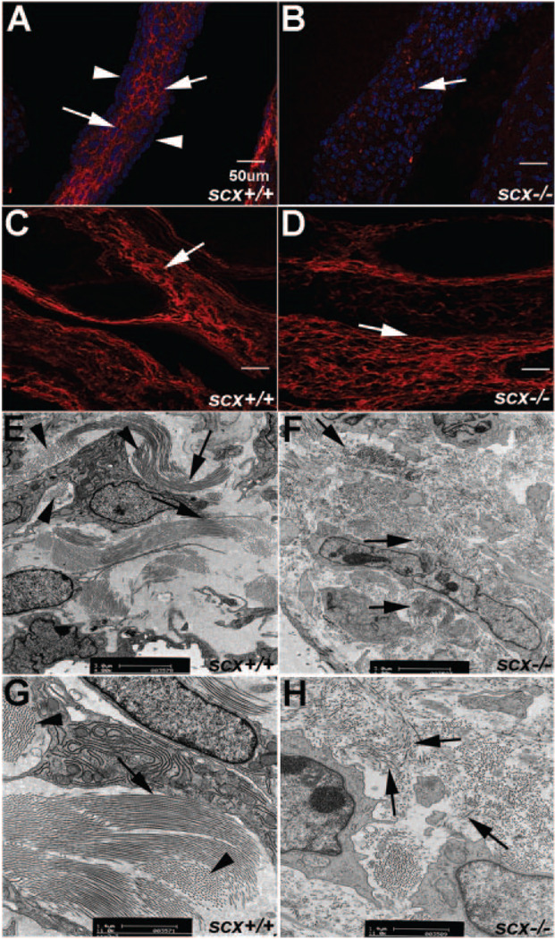
Figure 3. Expression of type XIV collagen is reduced and ECM organization is abrogated in valves from scX−/− mice.
A and B, Immunohistochemistry staining to show decreased type XIV collagen expression in heart valves from scX−/− mice (B) compared to scX+/+ mice (A) at E17.5. Arrows in A indicate type XIV collagen expression within the leaflet, whereas arrowheads show diminished expression on the leaflet surface. C and D, Type XIV collagen expression levels are comparable in vessels from scX+/+ and scX−/− mice. E through H, Transmission electron microscopy to determine collagen fiber organization in aortic valves from postnatal scX−/− mice, compared to scX+/+ mice. E and G, Long parallel collagen fiber bundles (arrows) and fibers running perpendicular (arrowheads) are observed in scX+/+ mice (arrows). F and H, Fragmented and disorganized collagen fibers are prevalent in scX−/− mice (arrows).

