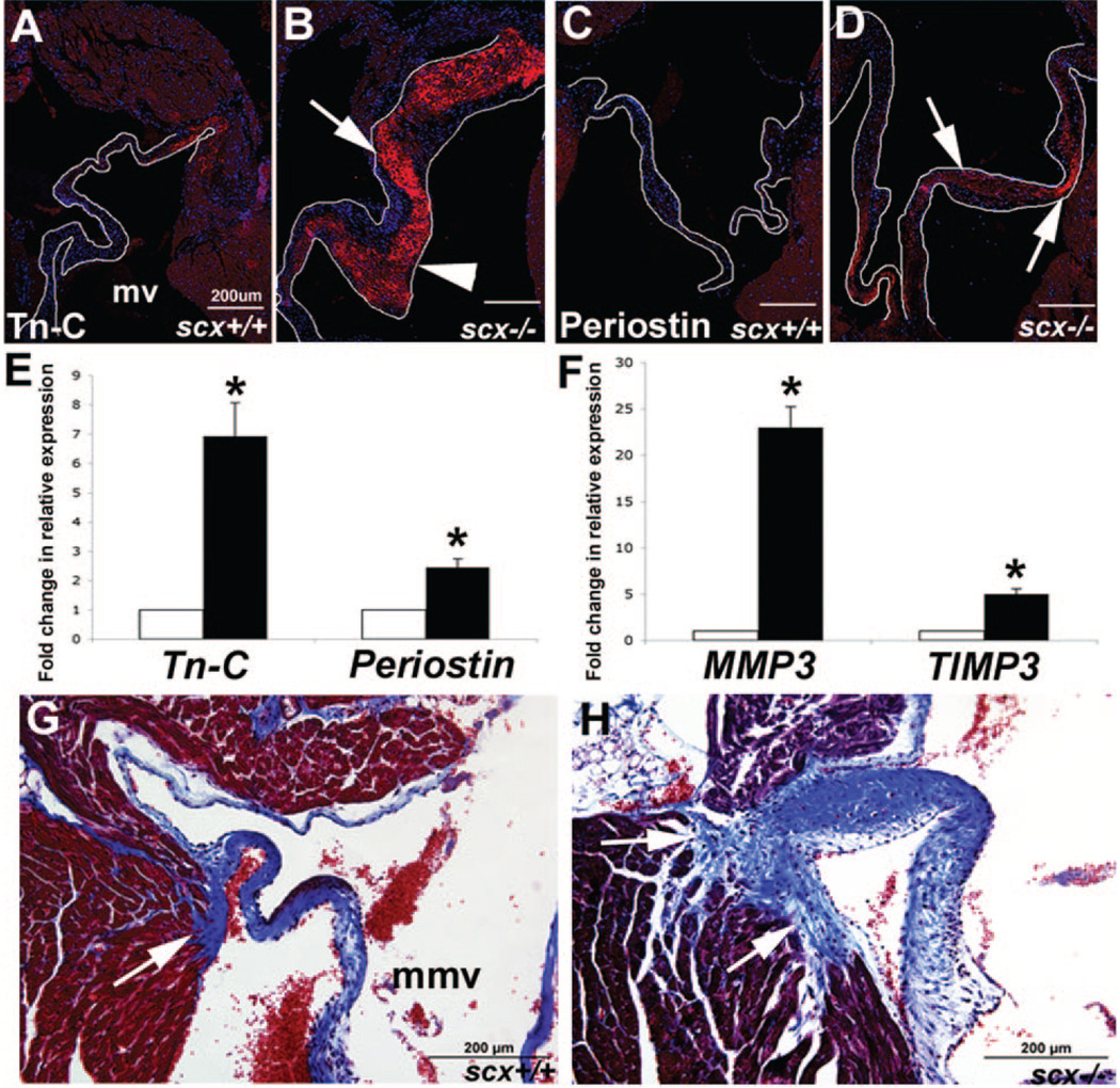
Figure 6. Increased ECM deposition and remodeling is observed in the AV region of juvenile scX−/− mice.
A through D, Immunohisto-chemistry was used to determine expression levels of tenascin-C (Tn-C) (A and B) and periostin (C and D) in scX−/− mice compared to scX+/+ mice at 2 months of age. Tn-C (A and B) and periostin (C and D) expression is significantly increased in valve leaflets from scX−/− mice. B, Note increased expression of Tn-C in ventricular regions of valve leaflet from scX−/− mice (arrowhead, compared to arrow). E and F, TLDA analysis shows significantly increased expression of tn-C and periostin at the transcript level in scX−/− mice compared to scX+/+ mice, as well as mmp3 and timp3. G and H, Trichrome staining to show collagen deposition (blue) in AV annular structures. Arrows indicate the posterior paraseptal annulus structure at the left AV groove. Note increased collagen deposition in scX−/− mice (arrows). mv indicates mitral valve; mmv, mural mitral valve.

