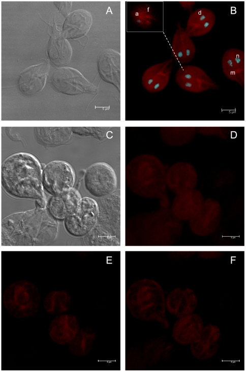
Figure 1. Localization of actin in Giardia lamblia trophozoites and cysts.
The cells were labeled using TRITC-phalloidin and analyzed by confocal microscopy. A, DIC image of trophozoites. B, Trophozoites stained with TRITC-phalloidin (d = ventral disc; m = median body; n = nuclei; and, f = flagella). C, DIC image of cysts. D, Cysts stained with TRITC-phalloidin. E and F, optical slices of D. The B inset represents an optical slice. The nuclei were labelled with DAPI.

