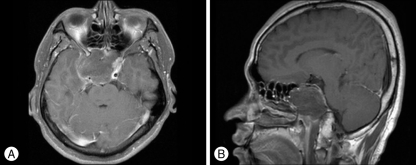
Fig. 2.
Preoperative magnetic resonance image (MRI) with gadolinium (Gd) enhancement. A : A lobulated and hypointense mass-like lesion in the axial image which fills the sphenoid sinus. It shows peripheral enhancement in the T1-weighted MRI scan with intravenous Gd injection. The size of mass is 5 cm×4.4 cm×5 cm. B : The pituitary gland and cavernous portion of internal cerebral artery are displaced upward to just below optic chiasm in the sagittal image.

