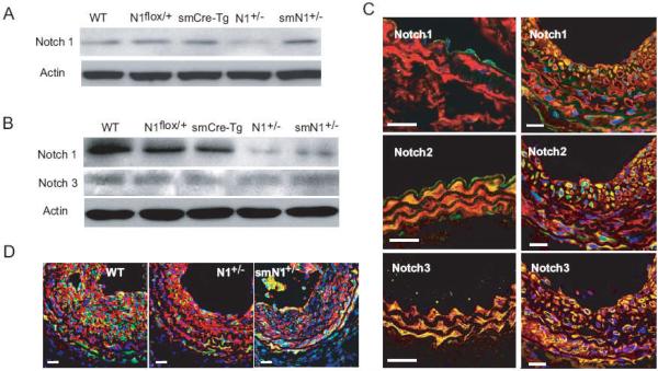
Figure 1. Expression of Notch receptors in endothelial and smooth muscle cells.
A. Western blot analysis shows that the expression of total Notch1 in EC isolated from hearts of WT, Notch1flox/+, smCre-Tg, N1+/-, and smN1+/- mice.
B. Western blot analysis shows that the expression of total Notch1 and Notch3 in SMC isolated from aortas of WT, Notch1flox/+, smCre-Tg, N1+/-, and smN1+/- mice.
C. Immunofluorescence staining of Notch1, Notch2, and Notch3 in uninjured carotid artery (left panel) and ligated injured carotid artery (14 days after ligation, right panel) of WT mice. Vessels were co-stained for smooth muscle actin (SMA). SMA is stained red; Notch receptors were stained green; and nuclei were stained blue (DAPI). Yellow indicates presence of both red and green staining. White bar was 20 μm.
D. Immunofluorescence co-staining of smooth muscle actin (SMA) with activated Notch1 (NICD). Red was SMA, green was NICD, and blue was DAPI. White bar was 20 μm.

