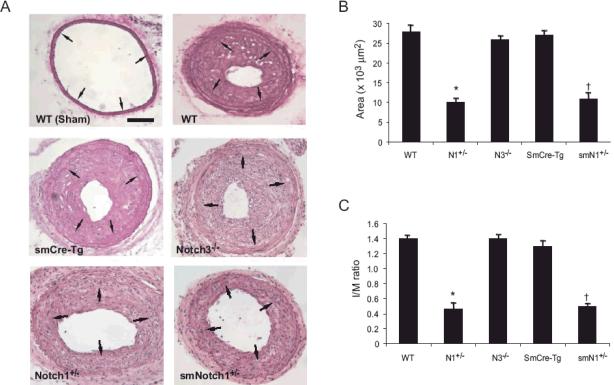
Figure 2. Neointimal formation in Notch mutant mice following vascular injury.
Carotid arteries were collected 28 days after the procedure. HE staining was performed on sections from WT, smCre-Tg, N1+/-, N3-/- and smN1+/- mice. Arrows indicate internal elastic lamina.
A. Representative sections are shown. Scale bar, 100 μm.
B. Quantitative morphometric analysis of intimal area in WT, smCre-Tg, N1+/-, N3-/- and smN1+/- mice. n=10 in each group, *; P<0.01, vs. WT; †; P<0.01, vs. smCre-Tg
C. Attenuated intima/media area ratio in Notch1 mutant, but not N3-/- mice compared with control mice. n=10 in each group, *; P<0.01, vs. WT; †; P<0.01, vs. smCre-Tg

