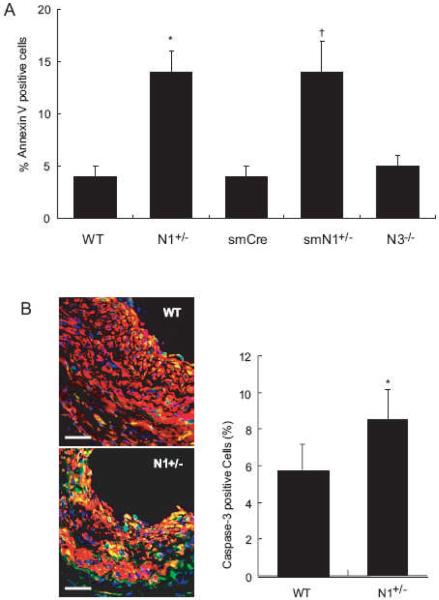
Figure 5. Apoptosis of SMC in Notch mutant mice.
A. H2O2-induced apoptosis of SMC. n=10, *; P<0.01, vs. WT; †; P<0.01, vs. smCre-Tg.
B. Left panel: Co-staining for SMA (red) and cleaved Caspase-3 (green) 14 days after ligation injury in WT and N1+/- mice. Right panel: Quantification of cleaved Caspase-3-positive cells in neointima from WT and N1+/- mice n=6, *; P<0.05, vs. WT

