Figure 3.

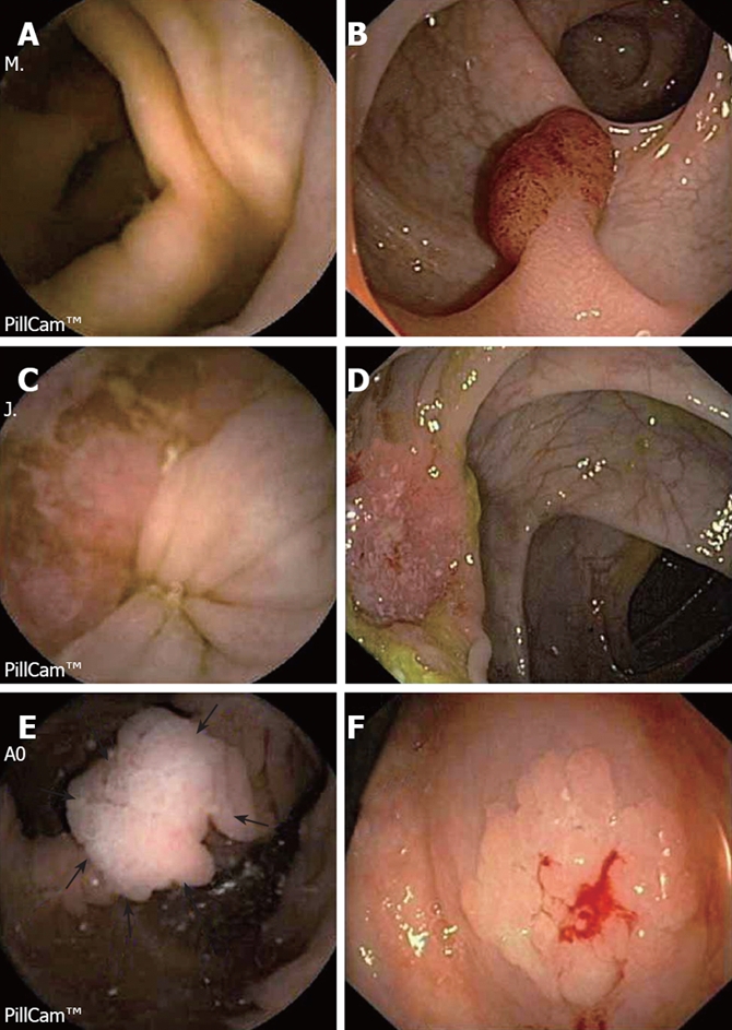
Images captured by the Pillcam™ Colon and conventional colonoscopy. A and B: Pedunculated polyp in the sigmoid colon; C and D: Ulcerated tumor in the transverse colon; E and F: Flat adenoma in the ascending colon.

Images captured by the Pillcam™ Colon and conventional colonoscopy. A and B: Pedunculated polyp in the sigmoid colon; C and D: Ulcerated tumor in the transverse colon; E and F: Flat adenoma in the ascending colon.