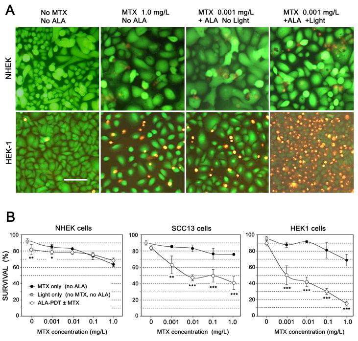
Fig 3. Preconditioning with MTX significantly enhances photocytotoxicity with ALA-PDT.

(A) Cell survival assays (using the fluorescent dyes, FDA and EB) following ALA-mediated photodynamic therapy with or without preconditioning with MTX. Monolayer cultures of cells (NHEK, top row; HEK1 cells, bottom row) were incubated for 72 h in the absence or presence of MTX, then were left unexposed or irradiated with intense blue light (395 nm, 4 mJ/cm2). At 24 h after irradiation, cells were incubated for 2 min with FDA and EB as described in Methods, then photographed using an epifluorescent microscope. Scale bar, 100 μm.
(B) MTX leads to PpIX accumulation and enhanced photodynamic killing of skin carcinoma cells, but has little effect upon normal keratinocytes. Cell cultures grown at the same time under similar conditions of confluency, were pretreated with MTX for 72 hr at the indicated concentrations, followed by 1 mM ALA for 4 hr. PpIX was visualized with confocal microscopy in some dishes to confirm PpIX induction (not shown), while other dishes were irradiated using a laser diode source. At 24 h post-irradiation, survival was analyzed using the FDA/EB assay; ~200 cells per field were counted to determine survival. Each data point represents mean ± SD of 3 micrographs. Open (white) circles: Cells pretreated with MTX, followed by ALA-PDT. Solid black circles: Cells given MTX pretreatment, but not exposed to ALA-PDT. Gray circle: Controls exposed to light only (no MTX, no ALA). (*) p<0.05; (*) p<0.01; (***) p <0.005.
