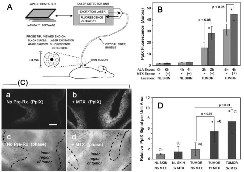
Figure 5. PpIX in chemically-induced skin tumors in mice.

Benign, low-grade papillomas were generated by chemical carcinogenesis on the dorsum of SKH-1 hairless mice, then pretreated with systemic MTX (or saline vehicle) for 1 or 3 days, followed by injection of ALA for 2h or 4 h prior to measurement of PpIX by noninvasive dosimetry (A, B), or by tissue biopsy (C, D). (A) Schematic diagram of the Aurora noninvasive fluorescence dosimeter. (B) Fluorescence signal is significantly increased in tumors from mice pretreated with MTX for 3 d, followed by ALA for the times indicated, as compared to normal skin (NL Skin) on the same mouse. Each bar, mean of 3 tumors (5 readings/tumor) ± SEM. (C) Mice were treated with/without MTX and with ALA, then euthanized and the tumors harvested and PpIX analyzed by confocal microscopy of frozen sections; typical PpIX signals (a, b) and corresponding tumor morphology (c, d) are shown. (D) Quantitation of the PpIX signal from digital confocal images, from tumors preconditioned with no MTX, 1 day of MTX, or 3 days of MTX prior to harvest. Shown are the mean ± SD of images from at least three independent tumors (numbers in parentheses).
