
Figure 3. A. NMR structure of RAP domains 1 (D1) (298), D2 (130), and D3 (131) showing the three helical bundle organization of each domain.
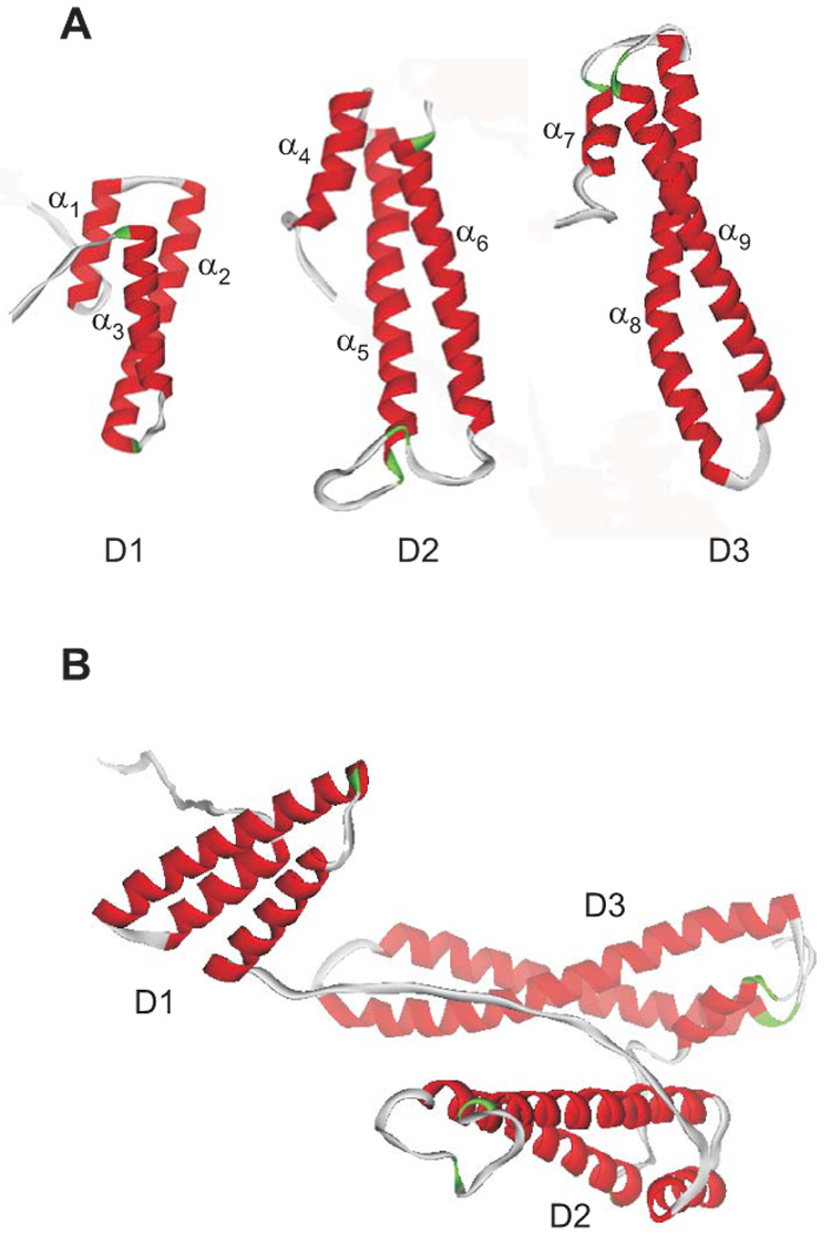
The helices are numbered as α1 – α9. B. Since the three domains of RAP are independent and do not interact, but are connected by long flexible loops, the protein is expected to adopt a variety of conformations in solution, one of which is shown.