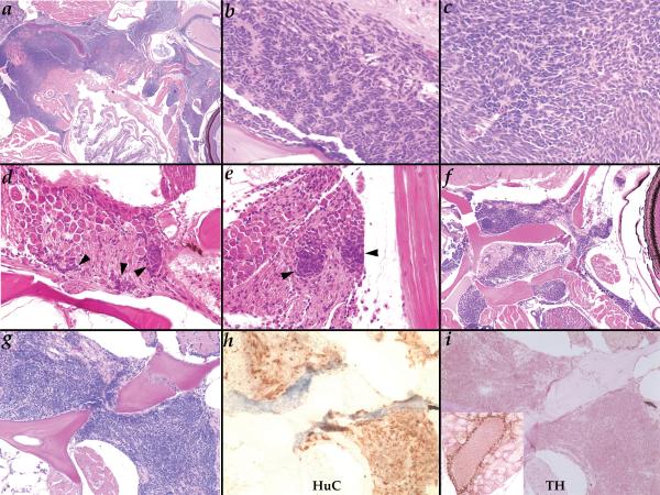
Figure 2.
Neuroblastoma-like tumors in Hag heterozygotes at two years of age. A. (40X) a very advanced tumor fills most of the head space between the esophagus and the skull and invades the body wall musculature; B-C. (400X) rosette arrangement of cells is occasionally observed in tumors (B) but more commonly cellular arrangement is more variable (C); D-E. (400X) very small neoplasias (black arrowheads) observed in the ganglia within the skull below the midbrain (D) or posterior of the ear (E); F. (100X) this tumor can be seen growing along the nerve running behind the eye as well as another below the skull; G-I. (200X) H&E (G), anti-HuC (H), and anti-TH (I) of the same tumor; inset in I shows TH staining of adrenal cells in the kidney on the same slide as a positive control for the antibody.

