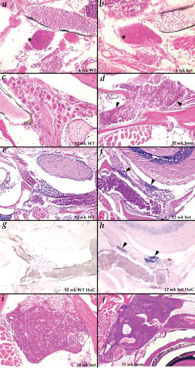
Figure 3.
Tumors begin as inappropriate maintenance of a normal developmental stage of putative neural precursors. A-B. (400X) at 4 weeks small cells resembling the tumor cells (asterisks) are seen in cranial ganglia of both wild type (A) and Hag (B) fish; C-D. (400X) at 12 weeks, only fully developed ganglion cells are observed in wild type fish (C), but the small precursor-like cells (black arrowheads) are still observed in most Hag mutants (D). E-H. (200X) the small cells seen uniquely in Hag mutants (black arrowheads) at 12 weeks (F) stain strongly for HuC mRNA by in situ hybridization (H); ganglion cells in wild type (E) and Hag mutants (F) stain weakly for HuC (G,H); I. (400X) tumor beginning to spread over entire ganglia in 24 week Hag heterozygote; J. (100X) tumor in 31 week homozygote growing within the skull and along nerves behind and above the eye.

