
Fig. 1.
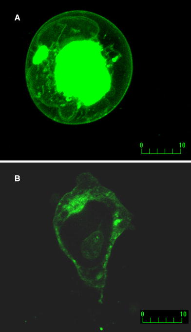
Confocal microscopy of the live tsA201 cells transfected with Nav1.5 + β1 (a) or Nav1.5 + β2 (b). Fluorescence of GFP linked to β subunit C-terminus at the cell membrane is evident for both β subunits. Optical slices were 0.5 μm (Zeiss Axiovert 100, Bio-Rad MRC 1024, excitation/emission wavelength 488/522 nm, laser power 10%)