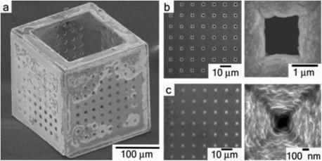
Figure 1.

a, Scanning electron micrograph (SEM) of a 200 μm three dimensional membrane with 10 μm pores on five faces and a large 150 μm pore on the sixth face. b, Zoomed-in detail of an array of 1.5 μm diameter pores on a single face of the membrane and a single micropore. c, Zoomed-in detail of an array of 100 nm diameter pores on a single face of the membrane and a single nanopore.
