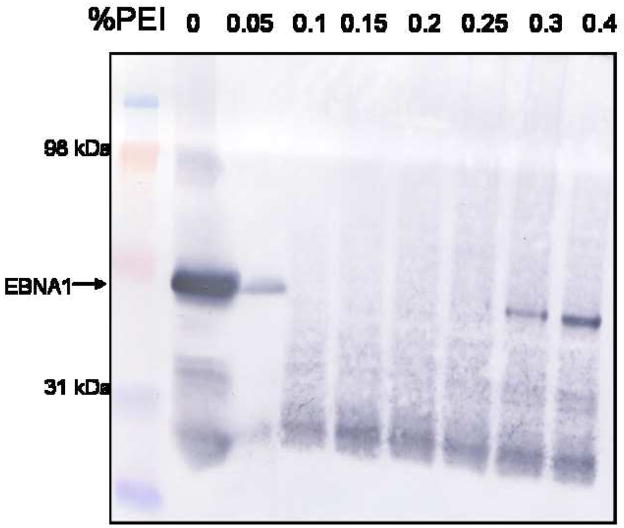
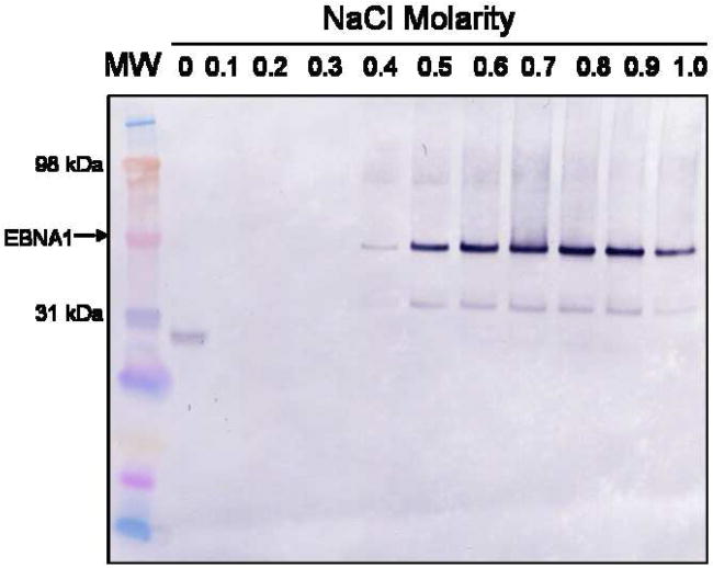
Figure 1. PEI precipitation of EBNA1.
A) PEI trial to determine the percentage of PEI necessary to precipitate EBNA1. Portions of whole cell extract were mixed with varying concentrations of PEI, the pellets were collected, and the supernatants were tested for the presence of EBNA1 by Western blot analysis with anti-EBNA1 mAb 1EB14. mAb 1EB14 binds in the N-terminal portion of EBNA1 suggesting that the EBNA1 fragment seen in each PEI sample lacks a functional DNA binding domain and is therefore not precipitated. B) Salt trials to determine the optimal concentration of NaCl to both wash the PEI pellet and elute EBNA1 from the pellet. 0.15% PEI pellets were washed with varying concentrations of NaCl, recovered by centrifugation, and the supernatants were tested for the presence of EBNA1 as described in A. MW is molecular weight markers.


