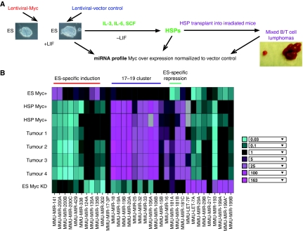
Figure 1.
miRNAs regulated by c-Myc in murine ES cells. (A) Scheme showing the experimental design for identification of miRNAs regulated by c-Myc in ES cells. c-Myc level was increased by lentiviral-delivered overexpression of c-Myc in ES cells (see Supplementary Figure S1). These ES cells, their differentiated haematopoietic stem/progenitor cell derivatives (HSP Myc+) and tumours after injection of HSP c-Myc+ cells into irradiated mice, were subjected to miRNA profiling. (B) Heatmap of miRNAs displaying significant regulation by c-Myc in ES cells and ES cell-derived differentiated cells or tumours. HSP: haematopoietic stem/progenitor cells derived by differentiation of ES cells; Tumours1–4: mixed T- and B-cell tumours derived by transplantation of Myc+ HSPs into irradiated mice. Note that results are shown for two HSP, and four tumour cell isolates independently derived from the same ES cell line. Bottom row shows miRNAs levels after c-Myc knockdown in WT ES cells. Inset: Fold change in miRNA copy number represents the ratio of miRNA level in ES cell in which c-Myc has been introduced (Myc+), or knocked down by c-myc shRNA (ES Myc KD), relative to lentiviral vector (see Supplementary Figure S1 for quantitation). Grey boxes represent no detectable signal.

