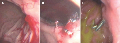
Figure 2.
Endoscopic view of a Dieulafoy’s lesion before and after endoscopic hemoclipping. A: Endoscopic view of a Dieulafoy’s lesion with active bleeding at the posterior wall of the proximal one third of the stomach. B: View after hemoclips application to bleeding site; bleeding has stopped. C: Endoscopic view of the same patient three months later.

