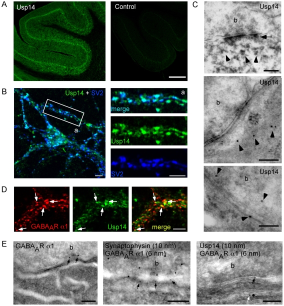
Figure 5. Usp14 and GABAAR α1 colocalize in cultured hippocampal neurons and cerebellar tissue slices.
(A) Immunohistochemistry on brain slices using Usp14-specific antibodies (PB+2, left) or without primary antibody (control, right). Further specificity controls are described by Crimmins et al. [9]. Scale bar 350 µm. (B) Coimmunostaining of cultured hippocampal neurons using Usp14- (PB+2, green) and SV2- (blue) specific antibodies. Turquoise puncta indicate partial colocalization of Usp14 and SV2 as a synaptic marker protein. Scale bars: 5 µm. (C) Electron microscopy analysis of DAB- (upper panel) or gold-labeled (middle and lower panel) Usp14 protein in Purkinje cells of mouse cerebellar slices using Usp14-specific antibodies (PB+2). Scale bars: 200 nm. (D) Coimmunocytochemistry of Usp14 [10] (green) and GABAAR α1 (red) reveals partial colocalization in neurites of cultured hippocampal neurons (yellow puncta, arrows). Scale bar: 5 µm. (E) Electron microscopy upon single-immunogold-labeling using antibodies specific for GABAAR α1 (left) or double-immunogold-labeling of GABAAR α1 with either synaptophysin- (middle) or Usp14 (PB+2, right). GABAAR α1 signals are found in close proximity to Usp14 signals at tubular submembrane organelles (b; synaptic bouton) of mouse Purkinje cells (PCs). Scale bars, left and middle: 200 nm. Scale bar, right: 100 nm.

