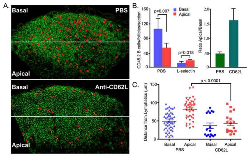
Figure 2.

Relocation of B lymphocytes within the lymph node follicle following blockage of lymphocyte ingress. (A) Location of transferred B cells. Twenty million CD45.2 B lymphocytes were transferred to recipient mice (CD45.1). Two hours later one group of mice (n=3) received 100 μg of CD62L antibody and the other group (n=3) received PBS. Eighteen hours after antibody injection the lymph nodes were harvested and immunostained for B220 and CD45.2. The CD45.2 positive cells are highlighted by red arrows. (B). A relative increase in number of transferred B cells in the apical region following CD62L antibody. The lymph node sections were divided into a basal region away from the lymphatics at the T-B boundary and an apical region close to the lymphatics. The number of CD45.2 B cells enumerated (left panel). The ratio between the numbers of cells in the apical and basal regions is shown (right panel). (C) Distance from lymphatics. The distances of the cells in the basal region from the subcapsular sinus and the distances of the cells in the apical region from the lymphoid cortical sinusoids were measured and plotted for the PBS and CD62L antibody treated mice. The statistical difference is between measured distances of the transferred B cells in the apical region of the inguinal lymph node from PBS versus the CD62L antibody treated mice.
