
Figure 3.
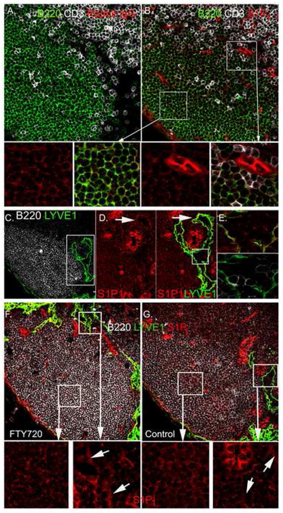
Location of S1P1 positive B lymphocytes within the lymph node follicle. (A-B) B220, CD3, and S1P1 immunostaining. Lymph node thin sections immunostained as indicated. Rabbit IgG is the control antibody (A) for S1P1 immunostaining (B). Magnified images of the white boxed regions are shown in the lower panels, the left two from deep in the follicle and the right two from the T-B border. (C) B220 and LYVE-1 immunostaining. Lymph node thin section immunostained as indicated. (D) S1P1 and LYVE-1 immunostaining around HEV and lymphatic. The left panel shows S1P1 immunostaining in the region outlined by the white box from part C. Yellow arrows indicate punctate staining of S1P1. The right panel shows S1P1 and LYVE-1 staining of the same region. (E) S1P1 immunostaining of cells in the lymphatics. Region outlined by white box in part D contains cells within lymphatic lumen. Upper panel shows LYVE- 1 (green) versus S1P1 (red) and lower panel shows LYVE-1 (green) versus B220 (white). (F-G) S1P1 immunostaining of lymph node sections from FTY720 treated and control mice. Mice were treated with FTY720 (1 μg/gm body weight for 18 hours or not). Lymph node sections (7 μM) were stained with antibodies against B220 (white), LYVE-1 (green) and S1P1 (red). White box areas are magnified and S1P1 staining is shown. Lymphatic endothelium staining is indicated (arrows).