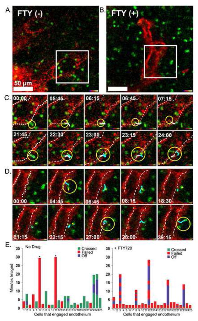
Figure 5.

B lymphocytes migration from the B cell follicle into adjacent lymphatic sinusoids is blocked by FTY720. (A,B) Single slice images (from supplementary movie 4 and 5) with indicated selected area were further analyzed below. Scale bar is 50 um. Similar protocol as was used as in figure 4. Selected white boxes were zoomed. (C,D) Image sequences from white box (from supplementary movie 4 and 5). Delineated lymphatic endothelium marked with doted white line. The yellow circle surrounds three penetrating cells in part C and the yellow circle surrounds two cells unable to penetrate barrier in part D. Time indicator shows the time of the snapshot image from 30 min movies. The dragon tail traces previous 6 time points. Representative of one of three experiments performed. (E) Individual transferred B cells that engaged a cortical lymphatic for a minimum of two minutes were observed during a 30 minute imaging period. The durations that 25 cells remained associated with the lymphatic endothelium were recorded from intravital TP-LSM imaging experiments performed with either control or FTY720 treated mice. The durations, which cells engaged the endothelium, but failed to cross, are indicated in red, while the durations, which cells engaged and passed, are indicated in green. If a cell disengaged from the endothelium and then re-engaged that duration is indicated in blue. Cells that remained bound to the endothelium for the duration of the imaging are indicated with an asterisk.
