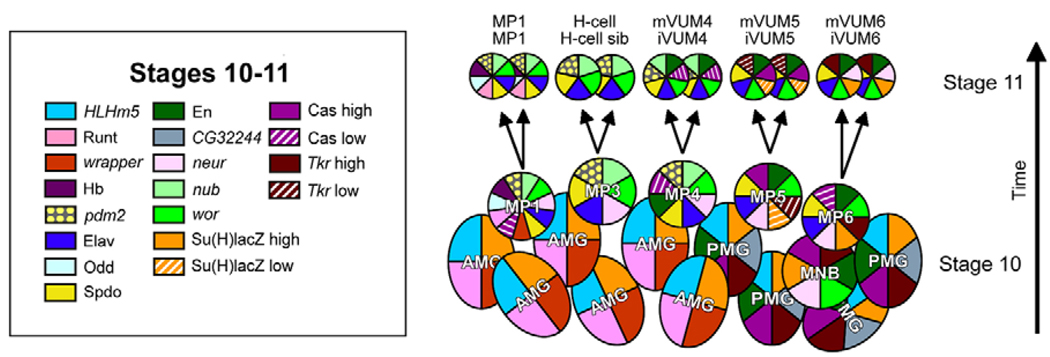
Fig. 1. Formation of midline precursors (MPs) and MP neurons in Drosophila.
Molecular map of stage 10 and 11 MPs and midline neurons (circles) and glia (ovals) shown in sagittal view. One segment is shown, with anterior to the left and interior (basal) at top. Each cell is depicted in terms of its pattern of gene expression as indicated by colors (the corresponding genes as listed on the left). The five MPs are shown at late stage 10, and the arrows indicate MPs dividing into their neuronal progeny at stage 11. The number of midline glia does not change appreciably from stage 10 to 11.

