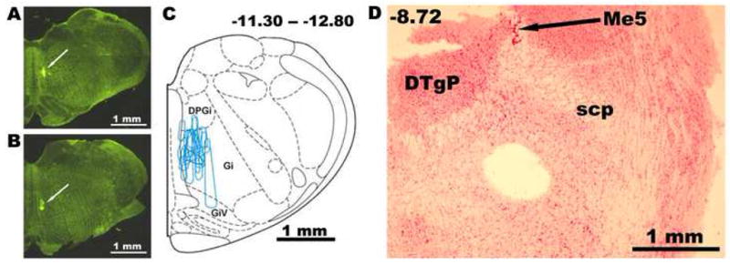
Figure 1.

FITC-labeled bead injection sites in the medial medullary reticular formation (A–C) and a typical location of a 600 μm micropunch of tissue taken from a pontine slice from a rat injected 5–6 days earlier with FITC-labeled beads (D). A and B: location of FITC deposit at two antero-posterior (AP) levels in one rat. C: injections sites as those shown in A and B from all animals superimposed on one standard cross-section located in the middle of the rostro-caudal span of the injected area that extended from AP-11.3 to AP-12.8 from bregma. Abbreviations: DPGi-dorsal paragigantocellular region; DTgP-dorsal tegmental region, pericentral; Gi-gigantocellular region; GiV-gigantocellular region, pars ventralis; Me5-mesencephalic trigeminal nucleus; scp- superior cerebellar peduncle.
