Figure 2.
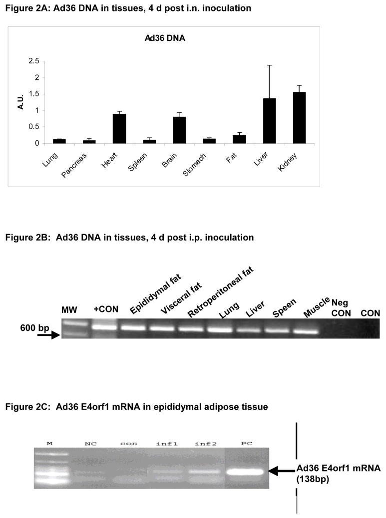
Figure 2A: Presence of Ad36 DNA in various tissues of rats infected by an intra-nasal inoculation. The quantities were determined by quantitative real time PCR.
Figure 2B: Presence of Ad36 DNA in various tissues of rats infected by intraperitoneal inoculation. The quantities were determined by nested PCR. MW: molecular weight ladder. Neg. CON: Water template, CON: Tissue from uninfected control animal.
Figure 2C: Ad36 E4orf1 mRNA in epididymal fat pad of two representative animals (inf 1 and inf2) from the Ad36 infected group. PC: Positive control mRNA from Ad36 infected A549 cells. NC: negative control water template. M: molecular weight ladder.

