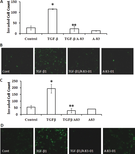
Figure 5.
TGFBR I inhibitor abolishes TGF-β1-induced invasion in EM42 cells and EECs. EM42 cells (A and B) and EECs (C and D) were treated with TGF-β1 (5 ng/ml) in the presence or absence of TGFBR I antagonist A 83-01 (5 µmol/l) prior to the transmesothelial invasion assay. A 83-01 was added at the same time as TGF-β. Data are presented as mean ± SEM (A and C), and representative images of invasion chambers are shown (B and D). (*P < 0.05 versus control; **P < 0.05 versus TGF-β1.)

