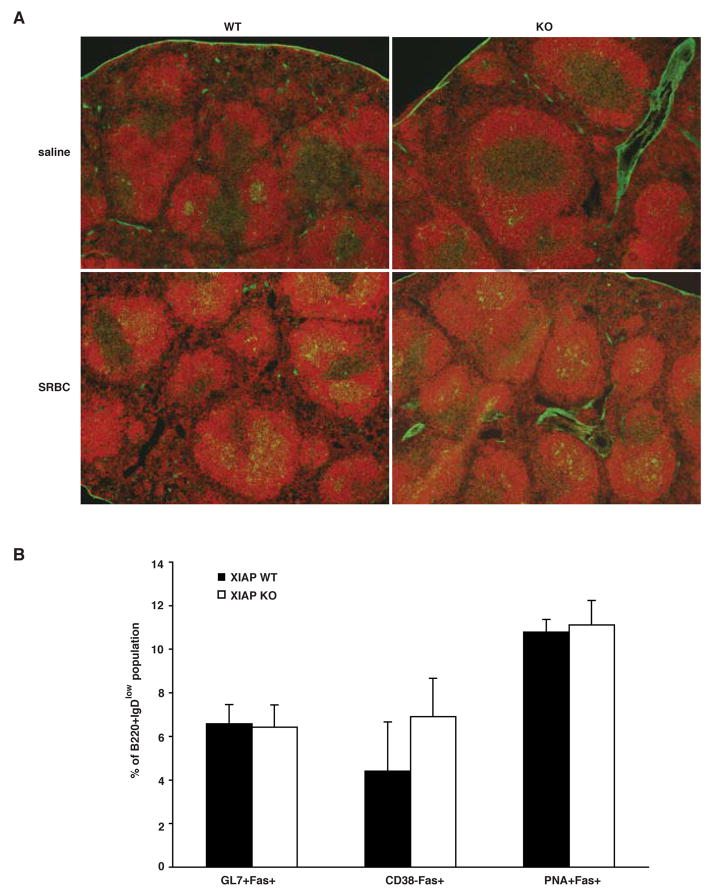
Fig. 4. Normal germinal center formation in XIAP KO mice.
(A) XIAP WT and KO mice were injected with either SRBC or saline and spleens were harvested 6 days later. Frozen sections were stained with PNA-FITC and anti-B220 and viewed on an Olympus microscope. (B) Splenocytes were harvested from mice treated in A and stained with antibodies to B220, IgD and Fas. Subsets were also stained with PNA or antibodies to GL7 or CD38 to specifically identify germinal center B cells. Data are representative of at least three individual mice, and error bars shown are standard error of the mean.

