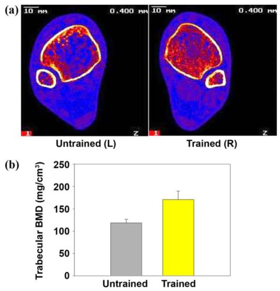
Figure 2.

(a) Peripheral quantitative computed tomography (pQCT) imaging of distal tibia of same subject in Figure 1. (Image is viewed from cephalad rather than caudad direction. In this subject, right leg underwent training.) Note extensive loss of trabecular lattice in untrained limb. L = left, R = right. (b) Distal tibia trabecular bone mineral density (BMD) for 3 subjects who trained >3 years was 30% higher in trained than untrained limbs (p < 0.05). Typical non-spinal cord injury BMD at this site is 250 mg/cm3. Source: Eser P, Frotzler A, Zehnder Y, Wick L, Knecht H, Denoth J, Schiessl H. Relationship between the duration of paralysis and bone structure: A pQCT study of spinal cord injured individuals. Bone. 2004;34(5):869–80 [PMID: 15121019]; Shields RK, Dudley-Javoroski S, Boaldin KM, Corey TA, Fog DB, Ruen JM. Peripheral quantitative computed tomography: Measurement sensitivity in persons with and without spinal cord injury. Arch Phys Med Rehabil. 2006;87(10):1376–81. [PMID: 17023249]
